Home
/ Dewinter Ekg : Patient 70yearold Male De Winter T Stock Illustration 1827946364, There is no anterior st segment elevation.
Dewinter Ekg : Patient 70yearold Male De Winter T Stock Illustration 1827946364, There is no anterior st segment elevation.
Dewinter Ekg : Patient 70yearold Male De Winter T Stock Illustration 1827946364, There is no anterior st segment elevation.. 11 minutes later, the chest pain was gone, suggesting spontaneous reperfusion (autolysis of thrombus). If the file has been modified from its original state, some details may not fully reflect the modified file. These patients are suffering occlusion myocardial infarction (omi) and require immediate reperfusion therapy. Right ventricular hypertrophy (mitral stenosis) old posterior mi. Ecg abnormality described by de winter et al.
Right ventricular hypertrophy (mitral stenosis) old posterior mi. T wave inversion ≥1 mm in at least two anatomically contiguous leads. The electrocardiogram (ecg) is one of the most useful diagnostic studies for identification of acute coronary syndrome (acs) and acute myocardial infarction (ami). It is rather rare (2% of cases). This study aimed to assess the prevalence and clinical characteristics of patients with this pattern.
Ecgs Life In The Fast Lane Flashcards Quizlet from o.quizlet.com De winter syndrome was initially described as a new ecg sign of occlusion of proximal left anterior descending (lad) coronary artery by robbert j de winter, niels j w verouden, hein j j wellens and arthur a m wilde, interventional cardiology group of the academic medical center, in 2008 as a letter to the editor. First reported by de winter in 2008, the de winter ecg pattern is an anterior stemi equivalent that presents without obvious st segment elevation. This file contains additional information, probably added from the digital camera or scanner used to create or digitize it. It is rather rare (2% of cases). Ischemia or posterolateral stemi) later: Ecg criteria for acute myocardial infarction: The de winter ecg pattern has been reported to indicate acute left anterior descending coronary artery occlusion and is often considered to be an 'st elevation myocardial infarction (stemi) equivalent'. There is no anterior st segment elevation.
N engl j med 2008;
He is also mildly diaphoretic. Secondary to occlusion of the proximal left anterior descending artery. The de winter ecg is sometimes seen in myocardial infarction with proximal lad occlusion. 1 among these, dwp is characterized by loss of r waves in the precordial leads. It is rather rare (2% of cases). This file contains additional information, probably added from the digital camera or scanner used to create or digitize it. Never trust the ecg computer interpretation, even if it says normal, because: Ecg abnormality described by de winter et al. Dewinter group is the premier provider of accounting, finance, & it professional staffing on an executive search, consulting & contract basis. The de winter electrocardiogram (ekg) pattern is a novel sign that indicates left anterior descending coronary artery (lad) occlusion in patients with chest pain. First identified in 2008 by dr. We aimed to investigate the morphology of the 'de winter ecg pattern' and evaluate the test chara … Ecg criteria for acute myocardial infarction:
The de winter ecg pattern has been reported to indicate acute left anterior descending coronary artery occlusion and is often considered to be an 'st elevation myocardial infarction (stemi) equivalent'. With atypical dewinter t waves. Secondary to occlusion of the proximal left anterior descending artery. This series shows the evolution of ecg changes in anterior wall m.i. T wave inversion ≥1 mm in at least two anatomically contiguous leads.
De Winter Sign Youtube from i.ytimg.com 1 this is the case of stemi equivalent patterns, such as hyperacute t waves, de winter's pattern (dwp), wellens syndrome, and posterior stemi. Ischemia or posterolateral stemi) later: It is rather rare (2% of cases). The de winter ecg pattern has been reported to indicate acute left anterior descending coronary artery occlusion and is often considered to be an 'st elevation myocardial infarction (stemi) equivalent'. Atrial flutter with variable block. These patients are suffering occlusion myocardial infarction (omi) and require immediate reperfusion therapy. Secondary to occlusion of the proximal left anterior descending artery. This study aimed to assess the prevalence and clinical characteristics of patients with this pattern.
With atypical dewinter t waves.
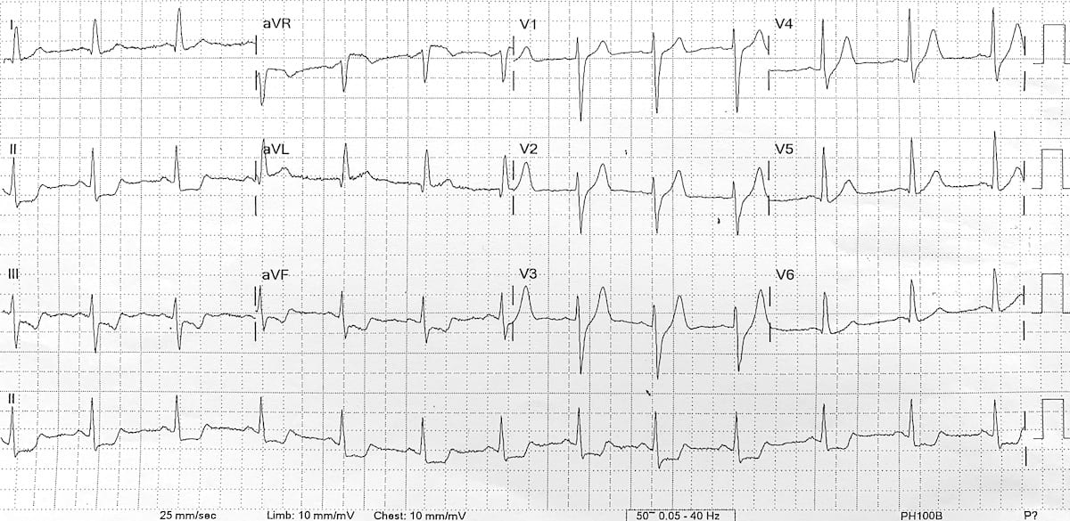
This series shows the evolution of ecg changes in anterior wall m.i. V1 already likely meets stemi criteria, and v2. This study aimed to assess the prevalence and clinical characteristics of patients with this pattern. The symptoms coupled with the shape of v1 and the dewinter changes as we advance through the precordial waves mean that at minimum this patient has acs, and repeat ekg's (every 15 minutes, possibly sooner depending on how he's responding to your treatment) and a call to cardiology are in order. There is no anterior st segment elevation. The de winter electrocardiogram (ekg) pattern is a novel sign that indicates left anterior descending coronary artery (lad) occlusion in patients with chest pain. Never trust the ecg computer interpretation, even if it says normal, because: This file contains additional information, probably added from the digital camera or scanner used to create or digitize it. The electrocardiogram (ecg) is one of the most useful diagnostic studies for identification of acute coronary syndrome (acs) and acute myocardial infarction (ami). It is rather rare (2% of cases). Atrial flutter with variable block. De winter syndrome was initially described as a new ecg sign of occlusion of proximal left anterior descending (lad) coronary artery by robbert j de winter, niels j w verouden, hein j j wellens and arthur a m wilde, interventional cardiology group of the academic medical center, in 2008 as a letter to the editor. Recognition of characteristic changes in an.
These patients are suffering occlusion myocardial infarction (omi) and require immediate reperfusion therapy. 1 this is the case of stemi equivalent patterns, such as hyperacute t waves, de winter's pattern (dwp), wellens syndrome, and posterior stemi. Ecg abnormality described by de winter et al. This study aimed to assess the prevalence and clinical characteristics of patients with this pattern. Dewinter group is the premier provider of accounting, finance, & it professional staffing on an executive search, consulting & contract basis.
De Winter T Wave Litfl Ecg Library Diagnosis from litfl.com Ischemia or posterolateral stemi) later: De winter syndrome was initially described as a new ecg sign of occlusion of proximal left anterior descending (lad) coronary artery by robbert j de winter, niels j w verouden, hein j j wellens and arthur a m wilde, interventional cardiology group of the academic medical center, in 2008 as a letter to the editor. With atypical dewinter t waves. The de winter electrocardiogram (ekg) pattern is a novel sign that indicates left anterior descending coronary artery (lad) occlusion in patients with chest pain. There is no anterior st segment elevation. First reported by de winter in 2008, the de winter ecg pattern is an anterior stemi equivalent that presents without obvious st segment elevation. V1 already likely meets stemi criteria, and v2. Secondary to occlusion of the proximal left anterior descending artery.
V1 already likely meets stemi criteria, and v2.
She was designated a cardiac alert from the field by paramedics. Ischemia or posterolateral stemi) later: With atypical dewinter t waves. Dewinter group is the premier provider of accounting, finance, & it professional staffing on an executive search, consulting & contract basis. We aimed to investigate the morphology of the 'de winter ecg pattern' and evaluate the test chara … The symptoms coupled with the shape of v1 and the dewinter changes as we advance through the precordial waves mean that at minimum this patient has acs, and repeat ekg's (every 15 minutes, possibly sooner depending on how he's responding to your treatment) and a call to cardiology are in order. The de winter electrocardiogram (ekg) pattern is a novel sign that indicates left anterior descending coronary artery (lad) occlusion in patients with chest pain. First reported by de winter in 2008, the de winter ecg pattern is an anterior stemi equivalent that presents without obvious st segment elevation. Recognition of characteristic changes in an. These patients are suffering occlusion myocardial infarction (omi) and require immediate reperfusion therapy. The de winter ecg pattern has been reported to indicate acute left anterior descending coronary artery occlusion and is often considered to be an 'st elevation myocardial infarction (stemi) equivalent'. If the file has been modified from its original state, some details may not fully reflect the modified file. A new ecg sign of proximal lad occlusion.
This study aimed to assess the prevalence and clinical characteristics of patients with this pattern dewi. Ischemia or posterolateral stemi) later: